All living cells are bounded by envelopes that protect them from the environment and confer their sizes and shapes. These shapes help cells to spatially organize their internal biological processes, allowing them to divide and faithfully segregate genetic material to each daughter. Yet, we still know very little about how cells obtain and control cell shape despite rapidly changing intra- and extra-cellular conditions, even in the arguably simplest and best understood organism: Escherichia coli. The problem is complex as it requires bridging scales between nanoscopic protein behavior and macroscopic cell shape and cell-cycle progression; this is where approaches from physics are required.
Here, I will present three vignettes that illustrate how my lab combines high-precision microscopy, microfluidics, physical modeling, and well-defined perturbations to reveal design principles and the relevant players of bacterial morphogenesis: 1) Cells constantly convert metabolites into biopolymers, notably protein, RNA, and DNA, which are densly packed inside the cell. To maintain the density of macromolecules within a permissive range, cells must therefore increase their volumes in coordination with the rate of biomass growth. We developed accurate label-free phase microscopy to measure cell mass and cell shape independently, which then allowed us to constrain and reveal design principles underlying this coordination. 2) Second, I will discuss how rod-shaped bacteria such as E. coli control rod-like geometry. Using single-molecule tracking and applying physical forces, we could show that different processes responsible for cell-envelope remodeling respond to different mechanical and geometric cues, which provide mechanical feedback for cell-shape control. 3) Finally, if time permits, I will discuss how bacteria decide when to divide into two. By following single-cell lineages in microfluidic chips over multiple generations, we could study temporal correlations between different cell-cycle processes. Those correlations then allowed us to build a stochastic model of cell-cycle control. We propose that not only one, but at least two independent processes must be completed before cells divide into two.
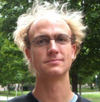
Sven van Teeffelen est professeur agrégé à l’Université de Montréal. Il est membre du Département de Microbiologie, Infectiologie et Immunologie de la Faculté de médecine. Sven a fait son doctorat en physique théorique de la matière molle à l’Université de Düsseldorf en Allemagne. Puis, comme postdoctorant à l’Université de Princeton, il a commencé à appliquer des approches expérimentales à des problèmes à l’interface entre la physique et la biologie, notamment concernant la forme et le cytosquelette des bactéries. En 2014, Sven a créé son laboratoire au sein de l’Institut Pasteur à Paris, ou il a continué à étudier la forme des bactéries et d’autres problématiques de la biologie physique des bactéries, en particulier la régulation du cycle cellulaire, celle de la densité intracellulaire, ou encore celle de l’intégrité de l’enveloppe bactérienne. Sven a intégré l’Université de Montréal en 2021. En 2019, Sven a reçu de l’Organisation européenne de biologie moléculaire (EMBO) le prix du jeune chercheur.

